X23D · AI Spine Navigation
Four X-rays. A full 3D spine, in flight.
X23D reconstructs a 3D lumbar spine from four intra-operative X-rays in 81 milliseconds per vertebra, fuses automated registration, and puts a tracked instrument on the overhead display before the first incision. Lower radiation, lower cost, no robotic gantry in the room.
Four numbers, straight off the cadaver bench.
Every figure here comes from the Luchmann et al. (2024) ex-vivo study and cited competitor literature. Nothing estimated, nothing rounded for effect.
Position, image, reconstruct, track, place.
The whole arc runs intra-operatively. Registration is automated through a titanium-bead bed module, so the step every legacy system fumbles simply does not exist here.
Position and calibrate
The patient lies on a table carrying a titanium-bead calibration module. That module is the single frame of reference for both the 3D model and the optical tracker, which is what makes the next step automatic.
1reference frame, sharedFour X-rays, four views
Four intra-operative X-rays are taken from four views, right before incision. No preoperative CT, no fluoroscopy loops during the case. This is where the radiation and cost savings begin.
4shots, then imaging is doneThe model builds itself
An AI trained by transfer learning traces spinal landmarks from pristine synthetic X-rays onto the real, blemished ones, effectively reversing the imaging process. It outputs a 3D lumbar model in about 81 milliseconds per vertebra.
81 msper vertebra, on the OR computerRegister, automatically
A third-party optical tracker reads the same titanium beads, so image and patient are already aligned the moment the model appears. No manual registration, no restart if the patient shifts on the bead-referenced frame.
0manual registration stepsInsert, watching the display
The surgeon watches the 3D model and the tracked instrument on the overhead display and places the pedicle screw along the planned trajectory. The whole case to this point has added little time over conventional 2D fluoroscopy.
1overhead display, the whole caseIt learns from spines it generates itself.
Train on generated X-rays, not real patients
Competing systems clean up their images with AI trained on radiology scans of real patients, the only images their trainers can legally acquire. That demand for real clinical data drives more scans, more radiation, more cost. X23D instead builds a 3D spine from a public CT database, renders thousands of artificial X-rays from every angle, and trains on those.
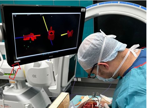
Trace clean landmarks onto blemished images
Transfer learning then feeds the AI both the pristine synthetic images and real, blemished X-rays, teaching it to carry the spinal landmarks from the perfect images onto the same landmarks in noisy ones. Given a new blemished image, it finds those landmarks and builds the 3D model, in effect reversing the process that created its training set.
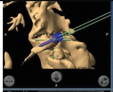
Where X23D wins, and where it does not.
Same procedure, lumbar pedicle-screw fusion, three systems. X23D takes two of three columns outright. The third is stated plainly below.
| System | X23D | 2D X-ray control | 3D nav + tracking |
|---|---|---|---|
| Overall breach rate | 21.00% | 24.00% | 25.60% |
| Clinically significant breach (Grade 2+) | 12.00% | 8.00% | 6.20% |
| Patient radiation (mSv) | 9.98 | 14.84 | 18.30 – 30.00 |
| Preoperative CT required | No | No | Yes |
| Robotic gantry in the room | No | No | Often |
Note. X23D and 2D control data from Luchmann et al. (2024). 3D-navigation breach data from Asada et al. (2024); 3D radiation data from Baumgart et al. (2023). Overall breach rate = screws deviating from the pedicle midline divided by screws placed. Clinically significant breach rate = Grade 2+ breaches divided by screws placed. Lower is better in every row.
The trade-off, stated plainly
X23D's clinically significant breach rate is higher. That is the whole story.
X23D's overall breach rate is the lowest of the three systems. But a Grade 0 or 1 breach is often asymptomatic, while a Grade 2+ breach can threaten a nerve root. On that measure, X23D sits at 12.00% against 6.20% for tracked 3D navigation. That is roughly double, and no marketing page can talk around it.
So the platform does not hide it. The clinical plan targets it directly, the positioning routes around it, and the roadmap treats bringing Grade 2+ under competitor parity as the single condition for full feasibility. Everything else on this page is already won.
A four-phase program to a 510(k) bridge.
X23D would combine a 510(k) clearance, enabled by a substantially equivalent device from Zeta Surgical, with PMA-grade controls reflecting the FDA's 2024 AI/ML action plan. Because X23D's algorithm is more advanced than Zeta's, more clinical information is needed for comparison.
Preclinical validation
Tests 3D-model accuracy under noise and poor positioning, and audits bias across 5,000 annotated X-ray sequences spanning BMI and age subgroups.
Pilot study
20–30 patients with varied spinal shapes. Goals: clinically significant breach under 7%, overall under 21%, radiation no higher than 10 mSv, with continuous algorithm monitoring.
Pivotal trial
160–310 fusion patients randomized to X23D or CBCT navigation. Primary endpoint: screw accuracy within 10% of CBCT. Secondary: radiation, insertion time, workflow interruptions.
FDA review
Algorithm frozen pre-trial, a Pre-Specified Change Control Plan governing future edits, and post-launch quarterly performance reports for the first two years.
Total program, approximately $17.2 million. Because the trial tests an AI algorithm, the model is frozen before enrollment and a change-control plan governs any post-market edit.
It does not fight the robots. It finds the rooms without one.
A Blue Ocean strategy: rather than compete head-to-head with established CT-based and robotic systems, X23D turns non-users of advanced navigation into adopters.
Who it is for
Experienced surgeons in 2D-fluoroscopy rooms
The target is older, rural surgeons who know 2D image-guided navigation but have never adopted 3D. Their free-hand experience offsets the major-breach rate (experienced surgeons breach less), and X23D's strong user experience eases adoption. They are not the academic centers already served by competitor devices.
The message that lands: less imaging, lower radiation, shorter operating time, at a fraction of a robotic system's footprint and cost.
What it removes and raises
Drop the gantry, keep the precision
X23D eliminates costly preoperative scans and large robotic systems, and raises radiation safety, overall breach avoidance, workflow speed, and access. The sustainable edge is a plug-and-play brand that harnesses ongoing AI advancement and network-driven learning, which late entrants struggle to replicate.
The condition for full feasibility: once the AI matures and Grade 2+ breaches fall below competitor parity, X23D's cost and radiation advantages make it the default, not the alternative.
Research status / X23D
A feasibility study.
A path to validation.
X23D has done no human clinical trials and is not being sold. This page reframes an academic technology-feasibility report as the product story it points toward. The full evidence, every citation, and the proposed FDA program live in the study behind the button.